fluoresence attached dna
Fluorescent nucleobases are chemically modified DNA and RNA analogues that not only retain their chemical and biological functionalities such as stacking base pairing and enzyme incorporation. In this case each of the nucleotide bases is labelled with a different fluorescent dye.
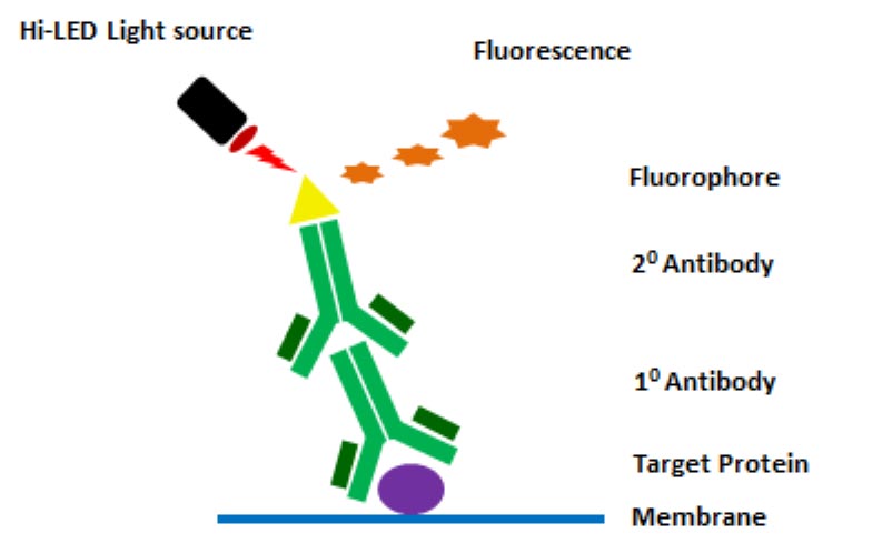
Fluorescence Applications Syngene
Especially interesting are DNA-PAINT combinations.
. Steady-state and time-resolved fluorescence of tetramethylrhodamine attached to DNA. Its four coding units A T C. The fluorescent bottlebrush polymer is then attached to an antibody via DNA hybridization resulting in an exceptionally bright immunofluorescent label.
A different coloured fluorophore is used for each of the reactions specific for the bases A C G and T. A depiction of the double helical structure of DNA. The nanoantennas are prepared by attaching one or two gold nanoparticles NPs to DNA origami structures which also incorporated docking sites for a single fluorescent dye next to one NP or in the gap between two NPs.
The simulations revealed that the dyes interact extensively with the G-quadruplex. The fluorescent DNA labeling can be carried out either in solution or on a reverse phase column. Fluorescence measurements involve exciting a dye molecule and then detecting the light that is emitted from the excited dye.
It takes advantage of the washing and separation properties of NPs and the structure-switch property of DNA aptamers resulting in fluorescence change of. The dye acts as the receiver part of. The nanoantennas developed by the team are synthesized from short segments of DNA with fluorescent dyes attached to certain parts.
Time-resolved fluorescence as well as steady-state absorption and fluorescence were detected in order to study the. Chen T 1 Fu L Zu L. The fact that DNA-PAINT can be readily realized with almost all conventional fluorescence microscopy modalities makes it particularly attractive.
Researchers discover that DNA naturally fluoresces. To better interpret such experiments classical and replica-exchange molecular dynamics simulations and fluorescence-lifetime measurements are used to understand the behavior of a range of Cy3-based dyes attached to the 3 end of G-quadruplex DNA. However their binding to DNA alters the structural and nanomechanical properties of DNA and thus affects also the associated biological processes.
1Department of Chemistry Beijing Normal University Beijing 100875 Peoples Republic of China. The next step is to label these probes by attaching one of a number of colors of fluorescent dyeDNA is composed of two strands of complementary molecules that bind to each other like chemical magnets. They are especially advantageous for the real-time monitoring of biochemical reactions and in vivo studies.
Amplified STR TechniqueInstrumentation Comments Reference. Our work builds on the initial discovery of fluorescence from few-atom silver clusters attached to a 12-base single-stranded DNA sequence11 We use six 19-base DNA oligomers to. DAPI fluorescence intensity increases if attached to DNA compared to its unbound state.
This is useful in many circumstances but sometimes it is necessary to incorporate a fluorophore closer to the DNA or RNA double helix without perturbing the helix. We have developed a method for the partial automation of DNA sequence analysis. Thetic DNA strands provide optically functional nanoele-ments with the desired small size sequence sensitivity and suitability for integration into DNA scaffolds.
Emission spectrum is broad and peaks at 461 nm. The emission color can be changed to orange or red by addition of. Correlation with DNA sequences.
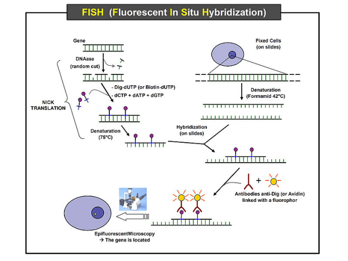
Fluorescent dyes are widely used for detecting and visualizing DNA molecules in many biotechnological applications. The fluorescence signal was quenched when carboxyfluorescein-labeled ssDNA was adsorbed by CoOOH. The technique relies on exposing chromosomes to a small DNA sequence called a probe that has a fluorescent molecule attached to it.
Fluorescence resonance energy transfer FRET is widely used in biomedical research as a reporter method. These procedures provide simple inexpensive methods of multiple DNA labeling and of introducing one fluorescent dye molecule per RNA as well as quantitative DNA fragmentation and incorporation of one label per fragment. In this case emission shifts to 500 nm.
It is excited by UV-light with a maximum at 358 nm. We measured the dependence of the fluorescence enhancement on NP size and number and compare it to numerical simulations. Fluorescence in situ hybridization FISH is a laboratory technique for detecting and locating a specific DNA sequence on a chromosome.
Oligonucleotides with a DNA backbone and one or several chromophore tags have found multiple applications as FRET probes. Fluorescent dyes can be attached to DNA molecules and modern automated methods for DNA sequencing make use of such fluorescent tagging. The simulations revealed that the dyes interact extensively with the G-quadruplex.
The probe sequence binds to its corresponding sequence on the chromosome. Fluorescence detection of the DNA fragments is accomplished by means of a fluorophore covalently attached to the oligonucleotide primer used in enzymatic DNA sequence analysis. These are called probes.
Fluorescent labels are commonly attached to the double helix at the end of a linker which places the fluorophore relatively far from the DNA bases. In the application to DNA typing with STR markers the fluorescent dye is attached to a PCR primer that is incorporated into the amplified target region of DNA. Abstract The turn-on fluorescence of pyrophosphate anion PPi was detected using DNA-attached cobalt oxyhydroxide.
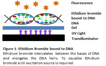
The intercalating dyes stabilize the duplex against thermal denaturation and show bright fluorescence in the green region of the spectrum. To better interpret such experiments classical and replica-exchange molecular dynamics simulations and fluorescence-lifetime measurements are used to understand the behavior of a range of Cy3-based dyes attached to the 3 end of G-quadruplex DNA. We have synthesized fluorescent DNA duplexes featuring multiple thiazole orange TO intercalating dyes covalently attached to the DNA via a triazole linkage.
A weak fluorescence can also be detected for RNA binding.
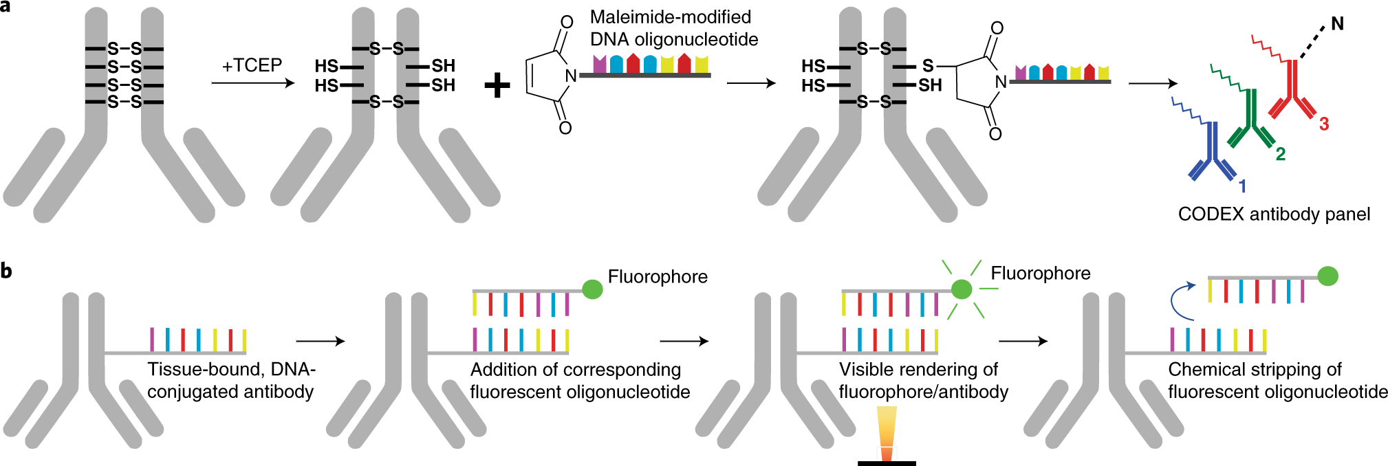
Codex Multiplexed Tissue Imaging With Dna Conjugated Antibodies Nature Protocols

The 10 Best Dna Dyes And Probes Biomol Blog Resources Biomol Gmbh Life Science Shop

Green Fluorescent Protein Gfp History Douglas Prasher
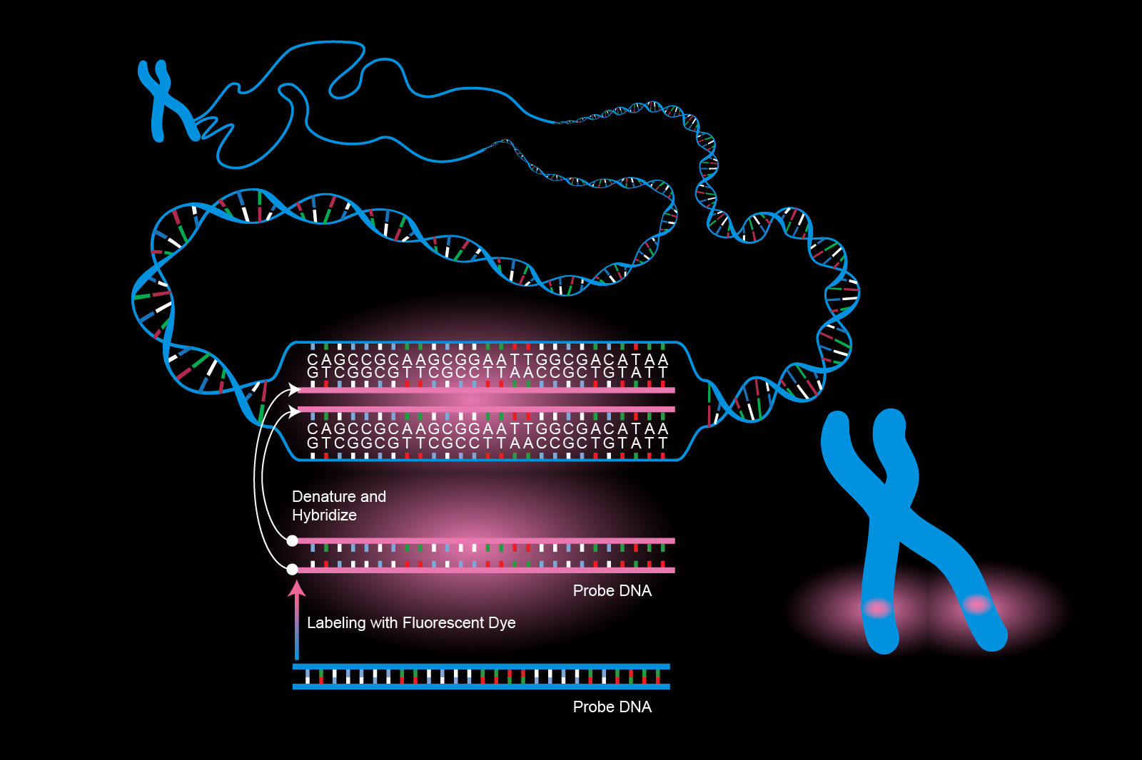
Fluorescence In Situ Hybridization Fish

Fluorescent Labeling What You Should Know Promocell

Fluorescence Resonance Energy Transfer In Near Infrared Fluorescent Oligonucleotide Probes For Detecting Protein Dna Interactions Pnas

Oligonucleotide Probe An Overview Sciencedirect Topics

Orientation Dependence In Fluorescent Energy Transfer Between Cy3 And Cy5 Terminally Attached To Double Stranded Nucleic Acids Pnas

Four Color Dna Sequencing By Synthesis Using Cleavable Fluorescent Nucleotide Reversible Terminators Pnas

Principles Of Fluorescence In Situ Hybridization Learn Science At Scitable
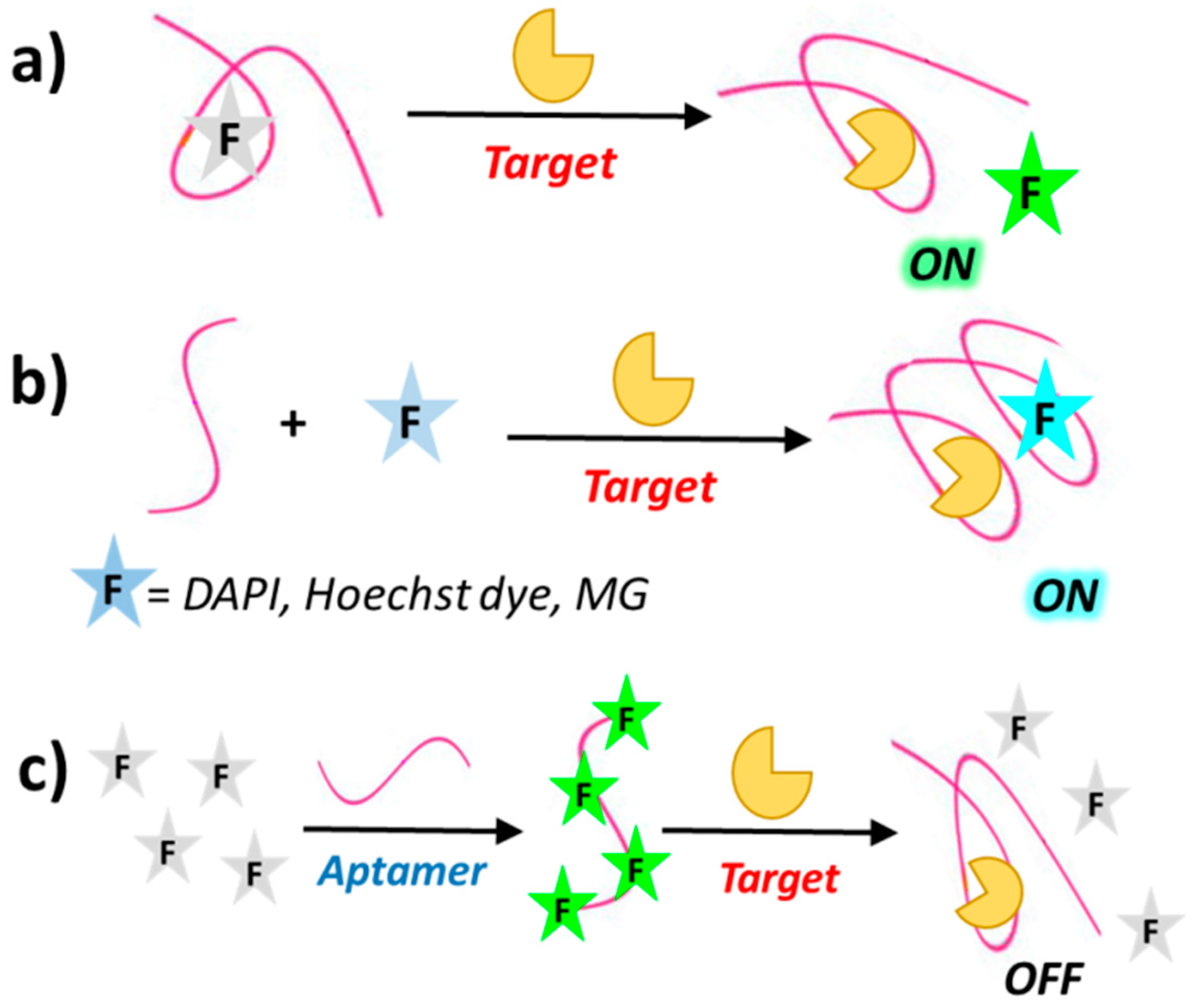
Cancers Free Full Text Fluorescence Sensing Using Dna Aptamers In Cancer Research And Clinical Diagnostics Html
Fluorescence Applications Syngene
2

Revealing The Competition Between Peeled Ssdna Melting Bubbles And S Dna During Dna Overstretching Using Fluorescence Microscopy Pnas
Fluorogenic Probes For Super Resolution Microscopy Organic Biomolecular Chemistry Rsc Publishing Doi 10 1039 C8ob02711k

6 Fam Reporter For Dna Probes Kilobaser Personal Dna Rna Synthesizer

Four Color Dna Sequencing By Synthesis Using Cleavable Fluorescent Nucleotide Reversible Terminators Pnas
Fluorescence In Situ Hybridization Fish Protocol Creative Biomart

Green Fluorescent Protein Gfp History Douglas Prasher


Comments
Post a Comment